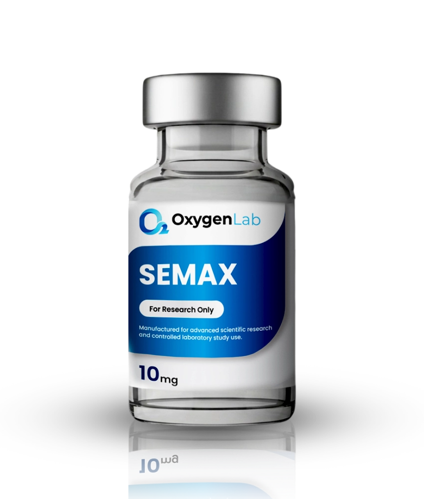
Research peptides usually arrive as a dry, freeze-dried powder sealed in a vial. Before that material can be used in an assay, it has to be dissolved in a known volume of liquid, and the moment you do that, one number starts to matter more than any other: the peptide concentration of the resulting solution.
If the concentration is off, every aliquot, serial dilution, and data point downstream inherits the same error. This guide covers the formula, worked examples, a reference table, unit conversions, and the most common mistakes.
If you just want the answer, our free peptide concentration calculator will do the arithmetic for you. Read on to understand what it is doing under the hood.
Why Peptides Ship Lyophilized
Most research peptides are supplied as lyophilized (freeze-dried) powder because removing water dramatically slows hydrolysis, oxidation, and microbial growth. A dry peptide can remain stable for long periods when kept cold and away from moisture and light. You can read more about the process in our article on what lyophilization is.
The trade-off is that lyophilized peptide reconstitution introduces a variable only the researcher controls: how much diluent is added.
The Core Formula
Peptide concentration after reconstitution is simply mass divided by volume:
Concentration (mg/mL) = Peptide mass in vial (mg) / Diluent volume added (mL)
From there, you can work out how much peptide is present in any volume you draw:
Peptide in an aliquot (mg) = Concentration (mg/mL) x Aliquot volume (mL)
And if you need a specific amount of peptide for an experiment, rearrange it:
Volume to draw (mL) = Target peptide amount (mg) / Concentration (mg/mL)
The errors creep in through units, not algebra.
Unit Conversions: mg, mcg, and mL
Mixing milligrams and micrograms is the most common source of thousandfold errors, so lock in these conversions first.
Mass
- 1 milligram (mg) = 1,000 micrograms (mcg or µg)
- 1 mcg = 0.001 mg
- 1 gram (g) = 1,000 mg
Volume
- 1 milliliter (mL) = 1,000 microliters (µL)
- 0.1 mL = 100 µL
- 0.01 mL = 10 µL
Concentration
- 1 mg/mL = 1,000 mcg/mL = 1 mcg/µL
- 1 mg/mL = 100 mcg per 0.1 mL
That last line is a handy shortcut: multiply a mg/mL value by 100 to get mcg per 0.1 mL. A 2.5 mg/mL solution therefore contains 250 mcg in every 0.1 mL (100 µL).
Worked Examples
Example 1: 5 mg Vial + 2 mL Diluent
- Concentration = 5 mg / 2 mL = 2.5 mg/mL
- In mcg: 2.5 x 1,000 = 2,500 mcg/mL
- Per 0.1 mL (100 µL): 2,500 x 0.1 = 250 mcg
If your assay calls for 100 mcg of peptide, you would draw 100 / 2,500 = 0.04 mL, or 40 µL.
Example 2: 10 mg Vial + 3 mL Diluent
- Concentration = 10 mg / 3 mL = 3.33 mg/mL (rounded)
- In mcg: 3,333 mcg/mL
- Per 0.1 mL: 333 mcg
Repeating decimals invite rounding errors. When possible, choose a volume that gives a clean concentration (for a 10 mg vial, 2 mL or 4 mL gives 5 or 2.5 mg/mL).
Example 3: 2 mg Vial + 1 mL Diluent, Then a 1:10 Working Dilution
- Stock concentration = 2 mg / 1 mL = 2 mg/mL (2,000 mcg/mL)
- Take 100 µL of stock and add 900 µL of buffer for a total of 1 mL.
- Working concentration = 2 mg/mL / 10 = 0.2 mg/mL (200 mcg/mL, or 20 mcg per 0.1 mL)
A concentrated stock diluted into a working solution is standard practice in cell culture and biochemical assays, and makes low final concentrations easier to hit accurately.
Reference Table: Common Vial Sizes x Diluent Volumes
The table below shows resulting concentrations as mg/mL and mcg per 0.1 mL. Values are rounded to two decimal places where needed.
| Vial Size | 0.5 mL | 1 mL | 2 mL | 3 mL | 5 mL |
|---|---|---|---|---|---|
| 1 mg | 2 mg/mL (200 mcg) | 1 mg/mL (100 mcg) | 0.5 mg/mL (50 mcg) | 0.33 mg/mL (33 mcg) | 0.2 mg/mL (20 mcg) |
| 2 mg | 4 mg/mL (400 mcg) | 2 mg/mL (200 mcg) | 1 mg/mL (100 mcg) | 0.67 mg/mL (67 mcg) | 0.4 mg/mL (40 mcg) |
| 5 mg | 10 mg/mL (1,000 mcg) | 5 mg/mL (500 mcg) | 2.5 mg/mL (250 mcg) | 1.67 mg/mL (167 mcg) | 1 mg/mL (100 mcg) |
| 10 mg | 20 mg/mL (2,000 mcg) | 10 mg/mL (1,000 mcg) | 5 mg/mL (500 mcg) | 3.33 mg/mL (333 mcg) | 2 mg/mL (200 mcg) |
| 15 mg | 30 mg/mL (3,000 mcg) | 15 mg/mL (1,500 mcg) | 7.5 mg/mL (750 mcg) | 5 mg/mL (500 mcg) | 3 mg/mL (300 mcg) |
| 20 mg | 40 mg/mL (4,000 mcg) | 20 mg/mL (2,000 mcg) | 10 mg/mL (1,000 mcg) | 6.67 mg/mL (667 mcg) | 4 mg/mL (400 mcg) |
Figures in parentheses are mcg per 0.1 mL (100 µL). High concentrations near the top-left corner may exceed a given peptide’s solubility, so always confirm the material has fully dissolved.
For combinations not listed here, plug your numbers into the peptide mixing calculator, which returns mg/mL, mcg/mL, and the volume needed for any target amount.
Common Calculation Mistakes
1. Confusing mg and mcg
Entering “500” when you mean 500 mcg but the formula expects mg produces a result 1,000 times too large. Write units next to every number, every time.
2. Ignoring Net Peptide Content
The mass printed on a vial is typically the gross weight of the lyophilized material. That powder can include counter-ions (such as acetate or trifluoroacetate salts) and residual moisture alongside the peptide itself. Net peptide content is often meaningfully below 100%. If your work requires precise molar concentrations, check the purity and net content figures on the certificate of analysis and correct your calculation accordingly.
3. Rounding Too Early
Rounding 3.333 mg/mL to 3.3 mg/mL before several serial dilutions compounds the error. Round only the final answer.
4. Forgetting Dilution Factors
A working solution’s concentration is the stock concentration divided by the dilution factor. Label every tube with its actual concentration, not just “stock.”
5. Losing Material to the Vial Walls
Powder often clings to the cap or walls. Briefly centrifuge the sealed vial before opening so all material collects at the bottom.
Choosing a Diluent for In-Vitro Work
The right diluent depends on the peptide’s sequence and the downstream assay. General guidance from peptide handling resources published by major reagent suppliers (for example, the peptide solubility and handling guidelines from Sigma-Aldrich, Thermo Fisher Scientific, and Bachem) follows a similar logic:
- Sterile, high-purity water is the usual first choice for short or charged peptides.
- Basic peptides (net positive charge) often dissolve more readily in a dilute acidic solution, such as 0.1% acetic acid, before further dilution.
- Acidic peptides (net negative charge) may dissolve better in a dilute basic buffer, such as a small amount of ammonium bicarbonate.
- Hydrophobic peptides may require a small volume of an organic solvent such as DMSO or acetonitrile first, followed by slow dilution into aqueous buffer. Keep final organic solvent levels compatible with your assay; many cell-based systems tolerate only low percentages of DMSO.
- Buffers like PBS are common for working solutions, but salts can reduce solubility of some peptides, so it is often better to dissolve in water first and add buffer afterward.
Test solubility on a small portion of an unfamiliar sequence first. Add diluent gently down the vial wall and swirl rather than shake, which can promote foaming and aggregation.
Storage After Reconstitution
Once in solution, peptides become far less stable than in dry form. General lab handling practice recommends:
- Aliquot immediately. Divide the stock into single-use volumes so the main supply is not repeatedly warmed and cooled.
- Avoid freeze-thaw cycles. Each cycle can degrade or aggregate peptide in solution.
- Store cold. Short-term storage at 2-8 °C may be acceptable for some sequences, while longer-term storage of aliquots is typically at -20 °C or -80 °C.
- Protect sensitive residues. Sequences containing cysteine, methionine, or tryptophan are prone to oxidation; using degassed or oxygen-free solvents and minimizing headspace helps.
- Label thoroughly. Record peptide name, lot number, concentration, diluent, and date on every tube.
For detail on keeping unopened material stable, see our guide on how to store lyophilized peptides.
Putting It All Together
Use the Oxygen Lab peptide concentration calculator to double-check your numbers before you pipette, and browse our research peptide catalog for lyophilized materials supplied with third-party testing documentation.
Frequently Asked Questions
How do I calculate peptide concentration after reconstitution?
Divide the peptide mass in the vial (in mg) by the volume of diluent added (in mL). For example, 5 mg dissolved in 2 mL gives 2.5 mg/mL, which equals 2,500 mcg/mL or 250 mcg per 0.1 mL.
How do I convert mg/mL to mcg per 0.1 mL?
Multiply the mg/mL value by 100. A 1 mg/mL solution contains 100 mcg in every 0.1 mL, and a 2.5 mg/mL solution contains 250 mcg per 0.1 mL.
Does the amount of diluent change how much peptide is in the vial?
No. The total peptide mass stays the same; only the concentration changes. Adding more diluent produces a more dilute solution, so you need a larger volume to obtain the same amount of peptide.
Why might my measured concentration differ from my calculated concentration?
Stated vial mass usually reflects gross weight, including counter-ions and moisture. Net peptide content, incomplete dissolution, and pipetting error all contribute. Check the certificate of analysis for net content and purity.
What is the best diluent for reconstituting research peptides?
Sterile high-purity water is the usual starting point. Basic peptides may dissolve better in dilute acetic acid, acidic peptides in a mild basic buffer, and hydrophobic peptides in a small amount of DMSO before dilution into aqueous buffer. Always match the diluent to your assay requirements.
For research use only. Not for human or veterinary use.